NEWS

Nuclear Medicine for Heart Health with Myocardial Perfusion Imaging
Nuclear medicine uses small amounts of radioactive materials to diagnose and treat a wide range of conditions.
It’s been revolutionary for heart health, providing doctors with detailed images of a heart’s function and blood flow. Managing heart health is vital for preventing serious conditions like coronary artery disease and heart attacks.
You may have heard of Myocardial Perfusion Imaging (MPI). It’s a key part of modern heart monitoring, helping your doctor effectively assess and treat heart issues. In this article, we’ll delve into everything you need to know about Myocardial Perfusion Imaging, its benefits, and how to prepare for the test.
What is Myocardial Perfusion Imaging?
MPI is a non-invasive nuclear medicine test that evaluates the blood flow to your heart muscle.
It’s most used to diagnose coronary artery disease, assess the severity of heart conditions, and guide treatment plans.
MPI gives healthcare professionals incredibly detailed images that show how well blood flows through your heart during times of rest and stress.
How does it work?
MPI uses a small amount of radioactive tracer that’s injected into your bloodstream.
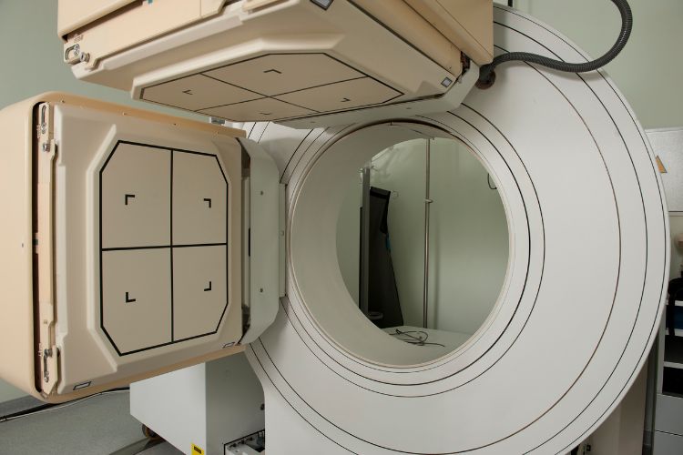
The tracer travels to your heart, where it emits gamma rays. A special camera, known as a gamma camera, detects these rays and creates images of your heart.
By comparing images taken at rest and after stress (induced by exercise or medication), your doctor can identify areas with reduced blood flow, which can be an indication of a blockage or another issue.
Types of MPI tests
There are two main types of Myocardial Perfusion tests: Stress MPI and Rest MPI.
Stress MPI involves exercising on a treadmill or stationary bike to increase your heart rate so the doctor can see the effects of physical stress on your heart. If you’re unable to exercise, you’ll be given medication that simulates the effects of exercise on your heart.
Rest MPI involves taking images of your heart while you’re at rest.
Your doctor will then compare the two sets of images to assess your blood flow and identify any abnormalities.

The Role of MPI in Diagnosing Heart Diseases
By evaluating the perfusion (blood flow) to the heart muscle, MPI can help your doctor identify areas with reduced perfusion, which can be a sign of a blockage or narrowed arteries.
During the test, both stress and rest images are taken to give insight into how well your heart functions under different conditions. This will help your doctor pinpoint the exact location and severity of any blood flow issues, leading to an accurate diagnosis and truly effective treatment planning.
What about other diagnostic tools?
MPI gives you and your healthcare team some powerful advantages, especially when compared to other diagnostic tools.
Unlike a standard ECG or stress test, MPI gives your doctor detailed images that show the structure and function of your heart. It can detect areas of poor blood flow that might not be apparent with other tests.
While an echocardiogram or cardiac MRI provide valuable information about your heart’s structure, MPI is particularly useful for assessing your blood flow and identifying any signs of Ischaemic Heart Disease (lack of blood or oxygen reaching your heart).
MPI is an essential tool in modern medicine, helping healthcare professionals diagnose and manage coronary artery disease for thousands and thousands of people.
Benefits of Myocardial Perfusion Imaging
Accurate and reliable
MPI is incredibly accurate and reliable in diagnosing heart conditions.
The scan gives your doctor detailed images, allowing them to assess your blood flow and identify areas of your heart muscle that might not be receiving an adequate supply of blood.
This precision helps your doctor diagnose conditions like coronary artery disease and determine how severe the blockages, if any, are.
Non-invasive
MPI is a safe and effective option for diagnosing heart issues. The procedure is non-invasive, which can be a significant advantage for a lot of patients.
It involves a simple injection of a radioactive tracer into your arm, followed by scans using a gamma camera.
Unlike invasive procedures, there are no surgical risks, and the process is generally easier for patients compared to other tests.
Early detection
With MPI, your doctor can detect the early signs of heart disease. It can be used to identify issues like reduced blood flow and potential blockages before they cause severe symptoms like chest pain or a heart attack.
Early detection gives your healthcare provider the opportunity for timely intervention and treatment.
By monitoring your blood pressure and heart function during the scan, MPI gives healthcare professionals valuable insights into your heart health.
Preparing for an MPI Test
Before the test
Before your MPI test, you should follow all the instructions provided by your doctor.
Typically, you’ll need to avoid caffeine for 24 hours before your appointment, including coffee, tea, and certain medications. You should tell your doctor about any medications you’re currently taking, as some may need to be paused.
If you are breastfeeding, you’ll need to discuss this with your doctor and may be required to take certain precautions after the test.
During the test
During your MPI test, you’ll receive a small dose of a radioactive tracer via an injection into your arm. A set of images will be taken while your heart is at rest.
You will then be asked to exercise on a treadmill or stationary bike to increase your heart rate, or you might receive medication that simulates exercise instead. The gamma camera will then capture detailed images of your heart at rest and under stress.
As mentioned above, the procedure is non-invasive and typically well-tolerated by patients, with minimal risk and discomfort.
Post-test considerations
After the test, you can usually go back to your normal routine straight away. Be sure to drink plenty of water to help flush the tracer from your body.
In terms of potential side effects, your doctor will discuss these with you, but they are rare and typically very mild.
Your results will be reviewed as soon as possible, and your doctor will follow up with you to explain the findings and discuss any further steps or treatments.
If you have any questions or concerns before or after your test, don’t hesitate to discuss them with your healthcare provider.
Safety and Risks of MPI
A common concern about MPI is the use of radiation. Read more.
While the procedure involves exposure to a small amount of radiation, the levels are extremely minimal and well within safe limits. Some patients might worry about potential reactions to the radioactive tracer, but adverse reactions are extremely rare.
The benefits of accurately diagnosing heart conditions and assessing your blood supply far outweigh any risks. MPI is a non-invasive exam, making it a safer alternative to invasive procedures.
Common misconceptions
One misconception is that MPI is only necessary for severe cases. In reality, it’s an extremely valuable tool for diagnosing conditions and assessing the severity of any blockages within your heart.
Another misconception is that the radiation used in the scan is harmful. The amount of radiation is carefully controlled and monitored for your safety.
Some patients also believe that MPI is only performed in a hospital, but actually, it can be done in an outpatient clinic like PRP, for your convenience and comfort.
It’s normal to fear the unknown, but it’s important to understand that MPI is a safe, effective, and non-invasive way to evaluate your heart health.
After the exam, the results and report will help your doctor create a tailored treatment plan to improve your heart health and quality of life.
The Future of MPI in Cardiology
Ongoing advancements in imaging technology and radiotracers make the future of MPI in cardiology look very promising.
Enhanced gamma cameras and medical software are making it possible to detect heart issues earlier and with even greater precision.
With these innovations, the healthcare community aims to improve the accuracy and efficiency of MPI, providing even more detailed images of the heart’s blood flow and function.
Here for your Health
For the best in diagnostic imaging, including MPI services, get in touch with PRP Imaging.
Our expert team is here to support your heart’s health needs with our top-of-the-line technology and compassionate care.