What are the 5 Medical Imaging Techniques?
Allowing doctors to look beneath the skin without a single incision, medical imaging technologies offer a window into the human body, providing images of organs, bones and arteries. These advanced techniques are pivotal in guiding today’s medical diagnoses and decisions, from revealing fractures to uncovering tumours.
As technology continues to evolve, there are various medical imaging techniques, each with its own diagnostic benefits.
Let’s explore the world of medical imaging and the five essential techniques that help doctors diagnose, treat and monitor various health conditions.
The 5 Medical Imaging Techniques Explained
X-ray Imaging
Discovered by Wilhelm Conrad Roentgen in 1895, X-ray is the oldest and one of the most commonly used medical imaging techniques. Roentgen’s accidental discovery occurred while testing if cathode rays could pass through glass. In a groundbreaking discovery, Roentgen discovered that these invisible rays could not only pass through glass but also penetrate human tissues and produce images of internal structures. This breakthrough was a medical revolution, enabling the first noninvasive diagnostic tool.
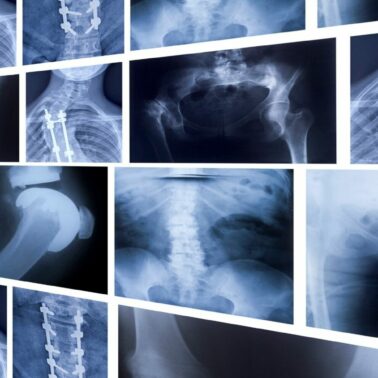
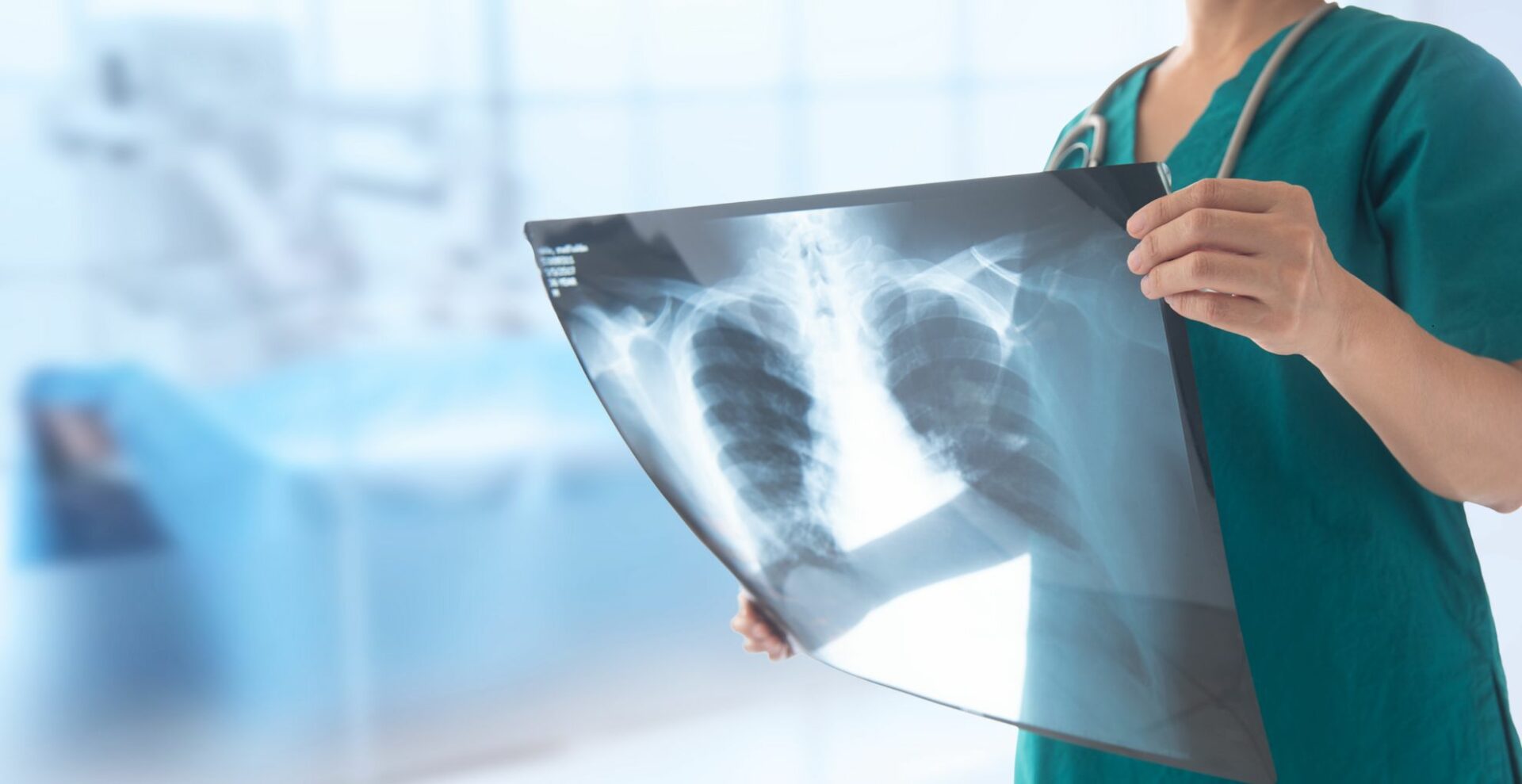
X-rays utilise electromagnetic radiation to penetrate the body and produce images based on the varying absorption of tissues. Dense tissues such as bone, absorb more radiation and appear white on the X-ray image, while softer tissues appear darker.
X-rays are beneficial for diagnosing bone fractures and lung conditions. They are also used by dentists for dental imaging and breast cancer detection or mammography.
Computed Tomography (CT)
Also referred to as a CAT scan, computed tomography (CT) is built upon X-ray technology to create detailed cross-sectional images of the body. CT scans provide three-dimensional views of organs and tissues by combining multiple X-ray images taken from different angles.
This imaging method is beneficial for diagnosing cancer, cardiovascular conditions, and trauma injuries as it provides a far more detailed image than traditional X-rays.

Iodine-containing contrast medium (ICCM) is a chemical used in X-ray imaging to enhance visibility of internal body structures, like blood vessels, organs, and joints. It can be injected into a vein or artery or ingested, helping radiologists get clearer images to diagnose conditions. ICCM is commonly used in angiograms, CT scans, and other specialised X-ray tests, and it interacts harmlessly with X-rays without producing radiation.
Magnetic Resonance Imaging (MRI)
Introduced into clinical practice in the 1980s, advanced medical resonance imaging revolutionised the healthcare field because of its ability to provide highly detailed images without the use of ionising radiation.
Instead, MRI employs powerful magnets and radio waves to generate detailed images of soft tissues. This technique is beneficial for diagnosing conditions in the brain, spinal cord, muscles and joints.
MRI scans are often used in neurology, orthopaedics, and cardiovascular imaging to provide clear, non-invasive views of internal structures. Contrast agents are used to enhance the clarity of MRI images. They help highlight specific areas of the body, making it easier for doctors to distinguish between normal and abnormal tissues. Common contrast agents include gallium, a substance that interacts with the magnetic field and improves the visibility of tissues or blood flow throughout the scan.
Ultrasound Imaging
Ultrasound uses high-frequency sound waves to create images of internal organs and tissues by measuring echoes and their bounce. This technique is safe, minimally invasive, and doesn’t involve radiation, making it a popular choice for monitoring pregnancy, heart conditions and abdominal issues. It can be used in 2-D, 3-D, and Doppler imaging to enhance visuals of blood flow.
Nuclear Medicine
Less commonly used than more standard methods like X-rays, CT scans, and MRIs, nuclear medicine involves using small amounts of radioactive materials, often referred to as tracers. These tracers can be injected, swallowed or inhaled to create images of the body.
Techniques such as PET scans and SPECT use gamma rays emitted by these tracers to detect cancer, heart disease and neurological disorders. Nuclear medicine is often integrated with other imaging techniques, such as PET–CT, for more comprehensive diagnostics.
Comparison of Imaging Techniques
Each medical imaging technique has specific advantages and is suited for diagnosing particular conditions. Here is a detailed comparison of the main imaging tests available, their applications, and their benefits and limitations.
1. X-ray Imaging
- Applications: Commonly used for diagnosing bone fractures, lung conditions and dental imaging.
- Advantages: Fast, widely available and excellent for visualising bones.
- Limitations: Uses ionising radiation, posing a risk with excessive exposure. It’s also been found to be less effective for imaging soft tissue.

2. Computed Tomography (CT)
- Applications: Used for diagnosing trauma, internal organ issues, and certain cancers.
- Advantages: These scans produce detailed cross-sectional images that are great for seeing soft tissues and bones and can quickly diagnose serious injuries.
- Limitations: CT involves more radiation exposure than standard X-rays. It’s more expensive, and it’s not ideal for either pregnant women or children.
3. MRI
- Applications: MRIs are ideal for brain, spinal cord, muscles and joint imaging.
- Advantages: No ionising radiation is used in these scans and they create highly detailed images of soft tissue.
- Limitations: MRIs are slower than CT scans and are less readily available in all areas. MRIs also have limitations when it comes to imaging bones.
4. Ultrasound
- Applications: These are commonly used in pregnancy and for monitoring heart conditions and abdominal issues.
- Advantages: Safe,minimally-invasive, and involve no radiation. Real-time imaging makes it suitable for guiding procedures like biopsies.
- Limitations: Limited ability to image deep tissues or bones and lack of ability to produce the high level of detail that CT or MRI scans can offer.
5. Nuclear Medicine
- Applications: Nuclear medicine is primarily used in oncology, cardiology, and neurology, such as PET and SPECT scans.
- Advantages: It offers functional imaging, allowing doctors to see how organs and tissues are functioning, which is critical in early disease detection.
- Limitations: Using radioactive materials involves higher complexity, cost, and radiation exposure risks.
Safety Considerations for Your Scan
While some imaging techniques, X-rays, CT, and nuclear medicine, involve exposure to ionising radiation, others, such as MRI and ultrasound, are completely radiation-free. CT and nuclear medicine scans have benefits for diagnosing life-threatening diseases, the radiation dose is closely managed, ensuring we use the lowest dose possible, especially for vulnerable populations like children and pregnant women. In some cases, contrast agents using MRI and CT scans may also cause allergic reactions, so patients should inform their healthcare provider of any sensitivities.

Future Trends in Medical Imaging
Undoubtedly, emerging technologies will continue revolutionising medical imaging in the healthcare industry. Innovations like AI and machine learning are improving image analysis, helping doctors, radiologists, and sonographers make faster and more accurate diagnoses.
There is also growth in the field of molecular imaging, which may allow for earlier detection of diseases at the most cellular level. These developments, combined with safer imaging modalities, promise a more precise and efficient future for medical diagnostics.
At PRP, we know how important it is to stay at the leading edge of technology and innovation. Our mission is to provide you and your family with the most appropriate and precise diagnostic imaging. Contact us today to book your appointment or to speak to one of our staff about your options.
