The Uses, Benefits and Applications of Nuclear Medicine
Modern medicine is an incredible thing in Australia, and recent advancements in the diagnostic techniques and therapeutic methods we employ here place our healthcare system as one of the most innovative and effective in the entire world. Diagnostic medicine procedures have gone from strength to strength, with improved accuracy and effectiveness with each research innovation.
Amongst these innovations is nuclear medicine, a non-invasive treatment that uses tracers and metabolism to deliver diagnostic precision. While the word “nuclear” comes with negative connotations for some people, nuclear medicine is one of the most effective methods for assessing the health and function of internal organs and other structures.
The rise of nuclear medicine has transformed oncology, and most of us will benefit from its techniques at some point in our lives. However, many people may be unclear as to what it is, how it is done, and what it is used for.
What is Nuclear Medicine?
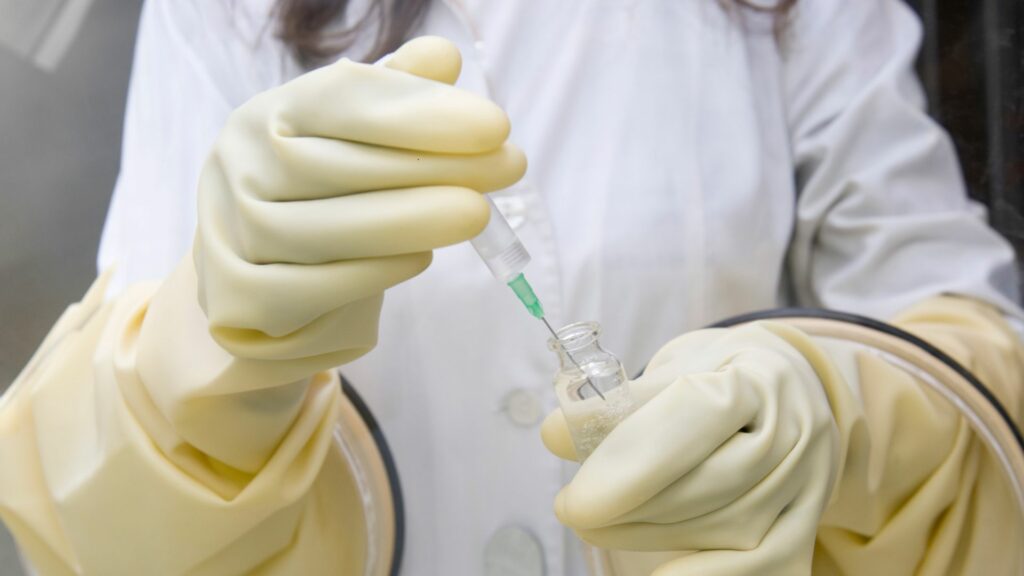
At its most fundamental, nuclear medicine is a form of medicine that uses a radiative tracer (or radiopharmaceutical) to examine organ and tissue function and to diagnose and treat disease. This tracer is typically inserted into the body via an injection. It then spreads around the body using the body’s metabolic pathways, spreading through organs and tissues. A specialised scan then captures the tracer’s radiation, revealing detailed images of the structure and function of organs and tissues in a way that traditional imaging simply cannot.
However, nuclear medicine is not just an effective diagnostic tool; it also has a role in therapy and treatment, and is an effective cancer treatment, combining the best of targeted therapy and radiation therapy. This means the radiopharmaceutical actively targets cancer cells, killing them with radiation but leaving healthy tissue relatively untouched. It can also be used to treat conditions of the thyroid, like hyperthyroidism or thyroid cancer, making nuclear medicine an incredibly important aspect of diagnosing and treating serious, life-threatening conditions.

Brief History of Nuclear Medicine
Although nuclear medicine may seem novel, it has seen widespread clinical use since the 1950s. By the 1960s, research had mapped the blood flow networks of the brain, and during the 1970s, most of the body’s organs could be mapped using nuclear medicine.
The American Medical Association officially declared nuclear medicine a specialised medical discipline in 1971, with single photon emission computed tomography (SPECT), which was developed in the 1980s, solidifying its place in routine diagnostic medicine. SPECT allowed the complete three-dimensional imaging of internal organs for the first time, which was later followed by the development of the first positron emission tomography scanner (PET), which allowed medical professionals to detect the emission of radioactivity from injected traces to assess and determine organ and tissue health and function.
These two innovative tomography techniques propelled nuclear medicine to the forefront of oncological care, and each year, procedures are becoming more effective, lower risk, and moving into the prediction and prognostic space, anticipating disorders and diseases before they fully develop.
How Nuclear Medicine Works
As its name implies, nuclear medicine harnesses radiation to diagnose and treat illnesses and diseases. At its core, radiation is a result of nuclear decay. Nuclear decay occurs when unstable atoms shed electrons, sending out energy in the form of waves and particles.
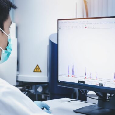
Radiopharmaceuticals contain elements that are undergoing nuclear decay and thus sending out energy. It is these waves and particles that can be detected during a scan.
Types of Radiopharmaceuticals
There are two main types of radiopharmaceuticals, those that help in detection, and those that help in treatment.
Diagnostic radiopharmaceuticals
Used in PET and SPECT, diagnostic radiopharmaceuticals emit energy that can be detected by scanning technology to compile a complete image of organ and tissue structure and function. These emissions allow for the creation of detailed, often three-dimensional, images that reveal organ and tissue structure and function.
Therapeutic radiopharmaceuticals
Used in radioactive iodine therapy and cancer treatment, therapeutic radiopharmaceuticals are radioactive drugs that seek out specific undesirable cells, such as cancer cells, before binding to them and killing them with radiation. They are also used to reduce the function of an overactive thyroid in hyperthyroidism.
Imaging Modalities in Nuclear Medicine
Nuclear medicine employs three primary types of imaging modality.
PET (Positron Emission Tomography)
Using a radioactive tracer, this imaging method is used to assess the metabolic or biochemical function of organs and tissues. Following normal metabolic processes, the tracer is drawn into organs and tissues and expelled again, and by scanning for the energy emitted by the tracer, we can see exactly how efficient (or inefficient) the metabolism of internal organs and tissues is.
SPECT (Single Photon Emission Computed Tomography)
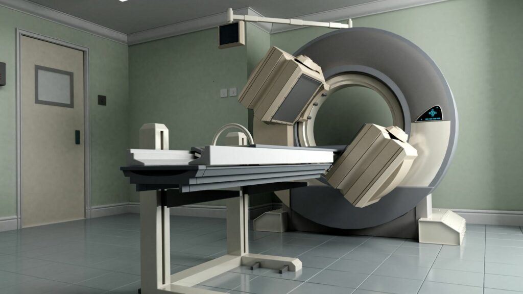
To develop three-dimensional models of organs, tissues, and other internal structures, SPECT uses another radioactive tracer, which is then captured by multiplanar nuclear medicine imaging technology. This provides an unparalleled 3D image of the internal body part, allowing in-depth study and analysis of its internal structures.
Hybrid imaging (e.g., PET/CT, SPECT/CT)
Sometimes, the use of hybrid imaging can give us a much more complete picture than just one method alone. In cases such as these, PET and SPECT can be paired with CT scanning. CT scanning is a type of X-ray that can provide two- and three-dimensional images of the entire body, so when paired with PET or SPECT, there is very little in the body that can escape such robust detection.

Diagnostic Applications of Nuclear Medicine
Common Uses of Nuclear Medicine in Diagnostics
Nuclear medicine is invaluable for the early detection of diseases such as cancer, cardiac conditions and neurological disorders—identifying abnormalities before symptoms become evident. This gives healthcare professionals an edge in preventative treatment by tackling the disease before it can spread throughout the body or progress beyond the point of effective treatment.
Functional imaging (e.g., brain activity, heart function) is another common reason to employ nuclear medicine’s diagnosis capabilities, as it gives a complete internal and external view of how organs and tissues are functioning during their metabolic processes.
Specific Examples of Nuclear Medicine Applications
Nuclear medicine is often employed to determine cancer staging and monitor its spread. The models developed through SPECT scanning can detect and measure tumour size and growth, while PET can assess if and by how much certain organs are working under additional strain. Over time, records of these scans can build a fuller picture of a cancer progression, including cancer metastasis.
While many people think of X-rays for this, nuclear medicine is also effective for scanning bones for fractures, especially compound fractures, providing a complete picture of the broken bone and where each piece is currently in the body.
Cardiac stress tests can also be conducted using nuclear medicine and have been done for decades. Scans can record heartbeats over a short period of time and assess the internal structure of the heart, including its chambers and valves.
Therapeutic Applications of Nuclear Medicine
Beyond diagnostic medicine and the monitoring of conditions and metabolic processes, nuclear medicine also has therapeutic applications and is used to treat diseases, including thyroid disorders and cancer. It does this by using radiopharmaceuticals to target and destroy harmful cells and tissues.
This form of radiation treatment is far more focused than traditional therapies that can affect and damage both cancerous tissue and healthy tissue. Radiopharmaceuticals do this by targeting specific cells, binding to them, and then delivering a lethal amount of radiation that kills the harmful cell.
Key Treatments Using Nuclear Medicine
One of the most common uses is in the treatment of thyroid disorders, such as an overactive thyroid or hyperthyroidism. Radioactive iodine therapy is used to treat this disorder, in which the patient drinks or swallows a capsule of radioactive iodine which then targets and destroys thyroid cells, reducing its overactivity back to normal, healthy levels.

Targeted radionuclide therapy is used in cancer treatment as an efficient and safe way to destroy cancer cells while leaving healthy tissue alone. To do this, the radionuclide is linked to a cell-targeting molecule so that it only seeks out and destroys cancer cells.
Bone pain palliation is a treatment that aims to reduce pain and maintain function in cancer patients who have metastasis in their bones and is considered one of the most effective treatment methods for relieving bone pain.
Emerging Therapies
New therapies are emerging all the time. One promising example is lutetium-177, which is being tested for the treatment of neuroendocrine tumours and metastatic prostate cancer.
Alpha-emitting radionuclides are also being tested for their efficacy in treating advanced cancers, effectively killing targeted cancer cells while ignoring healthy non-cancerous tissue.
Is Nuclear Medicine Safe?
The radiation doses used in nuclear medicine are incredibly minuscule, averaging between 0.3 and 20 mSv per procedure. In contrast, a CT scan delivers 7 mSv and flying on an aeroplane for a couple of hours exposes you to 0.035 mSv. While the levels in nuclear medicine are higher, there still isn’t a lot to worry about. Your healthcare provider will give you detailed information and advice post-procedure and most patients will pass all radioactive material in their urine in just a few days leaving no ill effects. However, care should be taken around family members, especially children, until all radiation is gone.
Minimising Risks of Nuclear Medicine
Medical facilities ensure patient and staff safety by using protective equipment for staff and using the minimum necessary amount of radioactive material to get a complete picture. It is also typically only used when other alternatives are not enough for diagnostic purposes.
The Australian Radiation Protection and Nuclear Safety Agency (ARPANSA) publishes a detailed code of practice relating to regulatory oversight and the global standards published by the World Health Organization are among the strictest in medicine.
Who Should Avoid Nuclear Medicine?
Precautions should be taken for pregnant or breastfeeding patients as even trace amounts of radiation can negatively impact the development and growth of a foetus or baby.
Technology in Nuclear Medicine
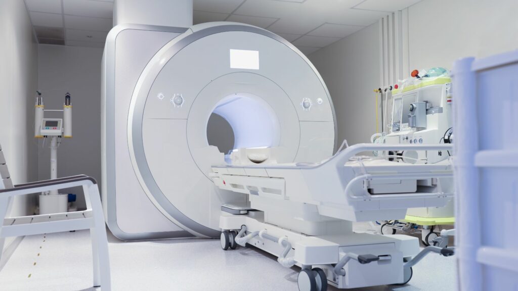
As the understanding of radiation by researchers developed, so did the technology to capture the emitted energy. PET scanners, SPECT cameras, and hybrid imaging systems all provide incredibly detailed models of internal structures and their metabolic processes. PET, SPECT, and hybrid all scan the patient in a similar way to CT scanning, where the patient lies down motionless and the scanning equipment moves across their body.
Advancement in Technology
Artificial intelligence (AI) is finding great traction in many industries and nuclear imaging is not different. Powerful AI algorithms assist in compiling and collating data to provide detailed imagery and diagnostic tools are also in development to assist in diagnostic, therapeutic, and workflow efficiency.
The development of new radiopharmaceuticals is also exciting, as different types of radioactive material will be suited for different functions. For example, lutetium Lu 177-dotatate can specifically target specific cancerous tumours, while ignoring normal, healthy tissue.
Nuclear Medicine vs. Other Modalities
Compared with other imaging techniques, such as CT, MRI, ultrasound, and X-ray, nuclear medicine offers information that no other technique can. While these four other modalities can also provide detailed and high-resolution imaging, only nuclear medicine can provide functional imaging, a unique benefit to this incredible diagnostic tool.
When Is Nuclear Medicine Preferred?
Nuclear imaging is the best option in cases where the function of organs is important to determine. For example, it can be incredibly useful to monitor the ongoing metabolic processes of the pancreas and other endocrine organs in cancer patients or to determine the function and efficiency of the heart in patients with heart disease. Ultimately, nuclear medicine works best when the clearest picture possible of internal structures is required to make a diagnosis.
What to Expect During a Procedure
Nuclear medicine may be an entirely new experience for most people, and you may not know what to expect at your appointment. It is best to wear loose, comfortable clothing to your appointment and feel free to share any concerns or ask any questions of your healthcare provider.
You will then be asked to sit down while the radioactive material is delivered. In some cases, like for the thyroid, this may be drunk as a liquid or swallowed as a capsule, but most cases require an intravenous injection for the best results.

Some radiopharmaceuticals will be taken up by the body immediately, and in these cases, your procedure may last no longer than 30 minutes from start to end. However, other radioactive tracers need more time to spread throughout the body. For these procedures, you may need to wait 2 or 3 hours after injection before the scan can be done.
After the allotted time, you will lie down on the scanning machine bed. Like an MRI or CT scan, it is important to remain as still as possible while the scan commences. A PET scan should take no more than 15–20 minutes to complete, while a SPECT scan can take 30–40 minutes.
Apart from the injection, the entire procedure should be entirely comfortable and pain-free. Reactions or side effects are incredibly rare, but your healthcare professional will be able to assist you with any symptoms.
Preparing for a Procedure
Preparation for a nuclear medicine procedure will depend on the precise nature of your procedure. Your doctor will advise if fasting is necessary and if there are any water intake restrictions. Some medications may also interfere with radiopharmaceuticals, so inform your doctor of any regular medication you are currently taking before scheduling your procedure.
Aftercare
Your body will be slightly radioactive after a nuclear medicine procedure and your healthcare provider will advise you on what to do depending on the specific procedure you have. It is advisable to drink a lot of water and try to urinate as much as possible in the first 24 hours as your kidneys will cleanse the radioactive material from your blood and pass it that way.
Avoid close contact with children or pregnant women as well, but it can be best to keep a little distance from everyone until most of the radioactive material has left your body, which is typically only a couple of days.
FAQs About Nuclear Medicine
“Is nuclear medicine safe?”
Yes, it is incredibly safe. The amount of radioactive material injected is incredibly minuscule and is usually completely flushed from the body in just a few days. However, we recommend that pregnant women or those who are breastfeeding consult with their doctor before undergoing nuclear medicine.
“How does it differ from X-rays or MRIs?”
Nuclear medicine differs from X-rays and MRIs as it allows us to see how effectively tissues and organs are functioning. In contrast, other forms of imaging only show a physical image with no indication as to the effectiveness of the organ’s function.
“How long does it take to get results?”
Given the enormous amount of information collected during imaging, results may take some time to fully collate. We will give you an indication of how long your results will take during your appointment.
Your Trusted Clinics for Nuclear Medicine Across NSW
The importance of nuclear medicine in modern healthcare cannot be understated. It is one of the most revolutionary technologies in healthcare and its benefits far outweigh its minuscule risks. As one of the most regulated and researched medicinal practices around the world, nuclear medicine is safe and effective, and recent advancements have and continue to broaden its scope to diagnose and treat a wide range of diseases and disorders.
PRP Imaging is proud to stand at the forefront of nuclear medicine in Australia as one of its most trusted and respected providers. With locations all through urban and regional NSW, we stand ready to provide safe and effective nuclear medicine services for all of your healthcare needs.
If you would like to learn more about the specific applications of nuclear medicine or schedule a consultation with us, please get in touch with our expert team today.
