
What is an Ultrasound
An ultrasound is a medical imaging technique that uses high-frequency sound waves to create images of the inside of the body. These sound waves are reflected off tissues, allowing doctors to visualise soft tissues and organs. This technique is also known as sonography.
Ultrasound is primarily a non-invasive procedure widely used for diagnostic purposes. It allows doctors to view organs, tissues, and blood flow in real-time. Specifically, ultrasound can be used to image the abdomen, pelvis (including obstetrics and gynaecology), blood vessels, and musculoskeletal soft tissues.
Whether you’re monitoring your pregnancy or investigating a health concern, ultrasounds give you and your healthcare provider a clear and detailed look inside your body. Ultrasound is especially valued for its safety and comfort for patients since it doesn’t use ionising radiation like X-rays or CT scans.
For those preparing for an ultrasound, understanding what to expect can help make the process smoother. Find key details below about ultrasounds to provide clarity on what they are, what’s involved during the procedure, preparation tips, and how to understand results.
How To Prepare For Your Ultrasound
Types of Ultrasound There are various types of ultrasounds designed to examine different areas of the body. Depending on the medical concern, an ultrasound may focus on the abdomen, pelvis, heart, muscles, joints, or specific organs such as the breasts. Here are some common types of ultrasound and their purposes:
Abdominal ultrasound
An abdominal ultrasound focuses on the organs within the abdomen, such as your liver, gallbladder, spleen, pancreas, and kidneys.
It’s commonly used to diagnose conditions like gallstones, liver disease, and kidney stones. This type of ultrasound can also help evaluate blood flow in your abdominal vessels.

Pelvic ultrasound
Pelvic ultrasounds are used to look at the organs in the pelvic area, including the uterus, ovaries, and bladder.
This type of ultrasound is particularly important in obstetrics and gynaecology, where it helps doctors monitor fetal development during pregnancy and diagnose conditions like ovarian cysts or uterine fibroids.
It’s important to note that pelvic ultrasounds can be carried out transabdominally (over the stomach) or transvaginally (through the vagina), depending on the area being examined. PRP Imaging have a large contingent of female sonographers to perform these types of ultrasounds, they will explain the process in detail and it will only be done with your consent.
Obstetric ultrasound
Obstetric ultrasounds are used during pregnancy to monitor the development and health of the fetus. These scans can confirm the pregnancy and the sex of the baby, estimate the due date, check fetal heartbeat, and detect abnormalities in the baby’s growth.
They provide crucial information about your baby’s growth, position, and overall well-being.
Breast ultrasound
Breast ultrasounds are used to examine breast tissue, often after an abnormal mammogram.
The procedure helps to identify cysts and solid masses. It’s an important tool in the diagnosis and monitoring of breast conditions, including cancer.
Cardiac ultrasound
A cardiac ultrasound, also known as an echocardiogram, is used to assess the heart’s structure and function. It helps diagnose heart conditions, evaluate blood flow, and monitor heart health.
This type of ultrasound gives doctors detailed images of the heart’s chambers, valves, and surrounding structures, making it incredibly important in cardiology.
Musculoskeletal ultrasound
Musculoskeletal ultrasounds focus on the muscles, tendons, ligaments, and joints. It’s commonly used to diagnose injuries like tears, sprains, and inflammation. This type of ultrasound is particularly useful in sports medicine and orthopaedics for evaluating soft tissue injuries.
There are various types of ultrasounds designed to examine different areas of the body. Depending on the medical concern, an ultrasound may focus on the abdomen, pelvis, heart, muscles, joints, or specific organs such as the breasts. Here are some common types of ultrasound and their purposes:
Abdominal ultrasound
An abdominal ultrasound focuses on the organs within the abdomen, such as your liver, gallbladder, spleen, pancreas, and kidneys.
It’s commonly used to diagnose conditions like gallstones, liver disease, and kidney stones. This type of ultrasound can also help evaluate blood flow in your abdominal vessels.
Pelvic ultrasound
Pelvic ultrasounds are used to look at the organs in the pelvic area, including the uterus, ovaries, and bladder.
This type of ultrasound is particularly important in obstetrics and gynaecology, where it helps doctors monitor fetal development during pregnancy and diagnose conditions like ovarian cysts or uterine fibroids.
It’s important to note that pelvic ultrasounds can be carried out transabdominally (over the stomach) or transvaginally (through the vagina), depending on the area being examined. PRP Imaging have a large contingent of female sonographers to perform these types of ultrasounds, they will explain the process in detail and it will only be done with your consent.
Obstetric ultrasound
Obstetric ultrasounds are used during pregnancy to monitor the development and health of the fetus. These scans can confirm the pregnancy and the sex of the baby, estimate the due date, check fetal heartbeat, and detect abnormalities in the baby’s growth.
They provide crucial information about your baby’s growth, position, and overall well-being.
Breast ultrasound
Breast ultrasounds are used to examine breast tissue, often after an abnormal mammogram.
The procedure helps to identify cysts and solid masses. It’s an important tool in the diagnosis and monitoring of breast conditions, including cancer.
Cardiac ultrasound
A cardiac ultrasound, also known as an echocardiogram, is used to assess the heart’s structure and function. It helps diagnose heart conditions, evaluate blood flow, and monitor heart health.
This type of ultrasound gives doctors detailed images of the heart’s chambers, valves, and surrounding structures, making it incredibly important in cardiology.
Musculoskeletal ultrasound
Musculoskeletal ultrasounds focus on the muscles, tendons, ligaments, and joints. It’s commonly used to diagnose injuries like tears, sprains, and inflammation. This type of ultrasound is particularly useful in sports medicine and orthopaedics for evaluating soft tissue injuries.
How to Prepare for a Abdomen Ultrasound? Please bring your referral, Medicare and Pensioner Health Care Cards with any previous imaging relating to the region being scanned.
Instructions on how to prepare for the test will be provided when you arrange the appointment. In general, there is a period of fasting for about 6 hours. Medications may be taken with a sip of water.
Please bring your referral, Medicare and Pensioner Health Care Cards with any previous imaging relating to the region being scanned.
Instructions on how to prepare for the test will be provided when you arrange the appointment. In general, there is a period of fasting for about 6 hours. Medications may be taken with a sip of water.
How to prepare for a Renal/Pelvic Ultrasound? Please bring your referral, Medicare and Pension Health Care Cards with any previous imaging relating to the region being scanned.
Instructions on how to prepare for the test will be provided when you arrange the appointment. In general, you will be asked to empty your bladder, drink and then hold 1-2 hours prior to the test.
Please bring your referral, Medicare and Pension Health Care Cards with any previous imaging relating to the region being scanned.
Instructions on how to prepare for the test will be provided when you arrange the appointment. In general, you will be asked to empty your bladder, drink and then hold 1-2 hours prior to the test.
What should I do the night before an ultrasound? The night before your ultrasound, it’s important for you to follow any specific instructions provided by your healthcare provider. Make sure to drink plenty of water to stay hydrated throughout your exam. On the day of the ultrasound, continue to follow any specific guidelines, such as avoiding certain foods or medications.
The night before your ultrasound, it’s important for you to follow any specific instructions provided by your healthcare provider. Make sure to drink plenty of water to stay hydrated throughout your exam. On the day of the ultrasound, continue to follow any specific guidelines, such as avoiding certain foods or medications.
What should I wear to my ultrasound appointment? Comfortable, loose-fitting clothing is ideal for your ultrasound appointment.
You might be asked to change into a gown, depending on the part of the body being examined. Avoid wearing jewellery or accessories that could interfere with the scan. Being comfortable will help you relax during the procedure.
Comfortable, loose-fitting clothing is ideal for your ultrasound appointment.
You might be asked to change into a gown, depending on the part of the body being examined. Avoid wearing jewellery or accessories that could interfere with the scan. Being comfortable will help you relax during the procedure.
What are the fasting requirements for an ultrasound? Many patients want to know if fasting is necessary, but this depends on the type of ultrasound you have. Abdominal scans often require fasting for about six hours to allow for clear images. Pelvic ultrasounds may require you to have a full bladder, so you might need to drink water and avoid urinating before the test. Our staff will give you specific preparation instructions based on your exam.
Many patients want to know if fasting is necessary, but this depends on the type of ultrasound you have. Abdominal scans often require fasting for about six hours to allow for clear images. Pelvic ultrasounds may require you to have a full bladder, so you might need to drink water and avoid urinating before the test. Our staff will give you specific preparation instructions based on your exam.
What is the arrival and check-in process for my ultrasound? Upon arrival at your local PRP Imaging clinic, you’ll check in at the reception desk, where our friendly staff will confirm your details and guide you to the waiting area. Be sure to bring your referral, Medicare or private health information, and any other relevant documents.
Our team is dedicated to ensuring you have a smooth and comfortable experience, taking care of all your needs throughout the process.
Upon arrival at your local PRP Imaging clinic, you’ll check in at the reception desk, where our friendly staff will confirm your details and guide you to the waiting area. Be sure to bring your referral, Medicare or private health information, and any other relevant documents.
Our team is dedicated to ensuring you have a smooth and comfortable experience, taking care of all your needs throughout the process.
What happens during the ultrasound procedure?
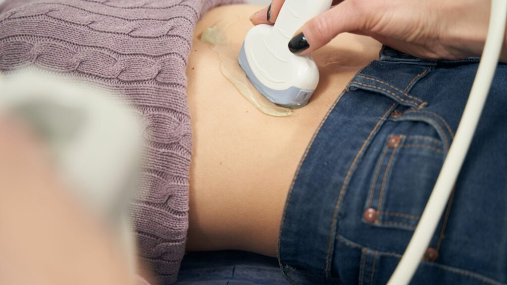
Understanding the examination process When you arrive for your ultrasound, a friendly technician will guide you to the examination room. You may be asked to change into a gown, depending on the area of your body being examined.
The technician will apply a clear gel to your skin — this gel helps the transducer, a handheld device, make better contact with your body to capture clear images. Once the gel is applied, the transducer is gently moved over the area being examined. For abdominal and pelvic ultrasounds, the technician will move the transducer over your skin to capture images of your organs. To get the best images can be very challenging and requires considerable time and concentration by the sonographer. With consent, an internal examination may be performed during a female pelvic ultrasound.
When you arrive for your ultrasound, a friendly technician will guide you to the examination room. You may be asked to change into a gown, depending on the area of your body being examined.
The technician will apply a clear gel to your skin — this gel helps the transducer, a handheld device, make better contact with your body to capture clear images. Once the gel is applied, the transducer is gently moved over the area being examined. For abdominal and pelvic ultrasounds, the technician will move the transducer over your skin to capture images of your organs. To get the best images can be very challenging and requires considerable time and concentration by the sonographer. With consent, an internal examination may be performed during a female pelvic ultrasound.
What will I see during the ultrasound? During the procedure, you’ll see black-and-white images on the screen. These images represent different parts of your body being examined.
For pregnancy ultrasounds, you might see your baby’s movements, heartbeat, and other details. The technician will take measurements and capture the necessary pictures to give a comprehensive view of your baby’s growth. Sometimes, for a more detailed view, a transvaginal ultrasound may be performed, where the transducer is gently inserted into the vagina to get closer images of the cervix and uterus.
During the procedure, you’ll see black-and-white images on the screen. These images represent different parts of your body being examined.
For pregnancy ultrasounds, you might see your baby’s movements, heartbeat, and other details. The technician will take measurements and capture the necessary pictures to give a comprehensive view of your baby’s growth. Sometimes, for a more detailed view, a transvaginal ultrasound may be performed, where the transducer is gently inserted into the vagina to get closer images of the cervix and uterus.
What do the technicians do during an ultrasound? The technician also referred to as a sonographer, will guide the transducer over your skin to capture various angles and measurements. They’ll remain with you throughout the scan and may ask questions about your medical history and the reason for the examination so that the optimal examination for your problem will be done.
To obtain clearer images, the sonographer may ask you to change positions during the procedure.
Throughout the test, your sonographer will make sure you’re comfortable and explain what they are doing. They’re there to answer any questions you have about the process.
The technician also referred to as a sonographer, will guide the transducer over your skin to capture various angles and measurements. They’ll remain with you throughout the scan and may ask questions about your medical history and the reason for the examination so that the optimal examination for your problem will be done.
To obtain clearer images, the sonographer may ask you to change positions during the procedure.
Throughout the test, your sonographer will make sure you’re comfortable and explain what they are doing. They’re there to answer any questions you have about the process.
How long does an ultrasound typically take? An ultrasound appointment typically lasts between 20 and 60 minutes, depending on the type and complexity of the scan. Check out the PRP service page for your scan for more information.
Some specific tests may take a shorter or longer amount of time. Your healthcare provider will be sure to disclose this before the procedure.
An ultrasound appointment typically lasts between 20 and 60 minutes, depending on the type and complexity of the scan. Check out the PRP service page for your scan for more information.
Some specific tests may take a shorter or longer amount of time. Your healthcare provider will be sure to disclose this before the procedure.
What are some important questions to ask my sonographer While our sonographers cannot discuss the result of the ultrasound with you, feel free to ask any questions during the procedure. You might want to ask about what they’re looking for and how the images will be used, and let them know about any concerns you might have.
Understanding the process can help reduce any anxiety you have and make the experience much more comfortable.
While our sonographers cannot discuss the result of the ultrasound with you, feel free to ask any questions during the procedure. You might want to ask about what they’re looking for and how the images will be used, and let them know about any concerns you might have.
Understanding the process can help reduce any anxiety you have and make the experience much more comfortable.
What should I do after my examination/ultrasound?
What to expect after the ultrasound After your ultrasound, you can expect to get back to your normal activities immediately. There are no known risks associated with ultrasound scans, making them a safe and convenient diagnostic tool.
The ultrasound technician will clean off any remaining gel from your skin, and you’ll be free to dress and leave the clinic as soon as you’re ready. If you had a full bladder for the scan, you’ll be able to use the facilities right away.
After your ultrasound, you can expect to get back to your normal activities immediately. There are no known risks associated with ultrasound scans, making them a safe and convenient diagnostic tool.
The ultrasound technician will clean off any remaining gel from your skin, and you’ll be free to dress and leave the clinic as soon as you’re ready. If you had a full bladder for the scan, you’ll be able to use the facilities right away.
How and when will I receive my results? The sonographer will capture and document the necessary images during your appointment. These images will be reviewed by a radiologist who will interpret the findings and complete a report.
Your results will typically be sent to your referring doctor within a few days. For more detailed scans, like a 3D ultrasound, the process may take a little bit longer.
Your doctor will discuss the findings with you and explain any identified issues or conditions. If any urgent issues are found, the clinic will contact you promptly.
The sonographer will capture and document the necessary images during your appointment. These images will be reviewed by a radiologist who will interpret the findings and complete a report.
Your results will typically be sent to your referring doctor within a few days. For more detailed scans, like a 3D ultrasound, the process may take a little bit longer.
Your doctor will discuss the findings with you and explain any identified issues or conditions. If any urgent issues are found, the clinic will contact you promptly.
Follow-up and additional testing requirements Depending on the findings from your ultrasound, your doctor may recommend follow-up appointments or additional tests. For example, if the ultrasound detects something unusual, such as a fluid buildup or abnormal size of an organ, further diagnostic tests may be necessary to understand the condition better.
In cases where birth-related conditions or other significant issues are identified, immediate follow-up actions will be advised by your doctor to ensure you receive the appropriate care and treatment.
Understanding your results and any required follow-ups will help make sure that you receive the best possible care. If you have any symptoms or concerns after your ultrasound, be sure to contact your healthcare provider for further guidance.
Depending on the findings from your ultrasound, your doctor may recommend follow-up appointments or additional tests. For example, if the ultrasound detects something unusual, such as a fluid buildup or abnormal size of an organ, further diagnostic tests may be necessary to understand the condition better.
In cases where birth-related conditions or other significant issues are identified, immediate follow-up actions will be advised by your doctor to ensure you receive the appropriate care and treatment.
Understanding your results and any required follow-ups will help make sure that you receive the best possible care. If you have any symptoms or concerns after your ultrasound, be sure to contact your healthcare provider for further guidance.
Your images and report from the ultrasound After your examination, the most pertinent images from your study will be available on the myPRP patient portal. A report, along with the images will be sent directly to your referring doctor. PRP will store digital copies of all studies on our secure database for comparison with any future examinations.
It is important that you return to your doctor with your examination results. Whether they are normal or abnormal, your doctor needs to know promptly so that a management plan can be formulated.
After your examination, the most pertinent images from your study will be available on the myPRP patient portal. A report, along with the images will be sent directly to your referring doctor. PRP will store digital copies of all studies on our secure database for comparison with any future examinations.
It is important that you return to your doctor with your examination results. Whether they are normal or abnormal, your doctor needs to know promptly so that a management plan can be formulated.
Ultrasound FAQs
Will the ultrasound procedure be painful? No, an ultrasound procedure generally isn’t painful at all. It’s a mostly non-invasive imaging technique that uses sound waves to create images of the inside of your body. You might feel slight discomfort from the pressure of the transducer, especially if you need a full bladder for your scan.
No, an ultrasound procedure generally isn’t painful at all. It’s a mostly non-invasive imaging technique that uses sound waves to create images of the inside of your body. You might feel slight discomfort from the pressure of the transducer, especially if you need a full bladder for your scan.
Can I eat or drink before my ultrasound appointment? Whether you can eat or drink before your ultrasound depends on the type of scan you’re getting. For pelvic ultrasounds, for example, you’ll need to drink plenty of water to fill your bladder.
Our team will give you specific instructions based on your exam.
Whether you can eat or drink before your ultrasound depends on the type of scan you’re getting. For pelvic ultrasounds, for example, you’ll need to drink plenty of water to fill your bladder.
Our team will give you specific instructions based on your exam.
What parts of the body can be examined using an ultrasound? An ultrasound can examine many parts of the body, including the abdomen, pelvis, heart, blood vessels, and musculoskeletal structures.
It’s commonly used for monitoring pregnancy, diagnosing conditions in both children and adults, and evaluating organs like the liver, kidneys, and thyroid.
If you need an ultrasound, our experienced team is here to provide a comfortable and professional imaging experience.
An ultrasound can examine many parts of the body, including the abdomen, pelvis, heart, blood vessels, and musculoskeletal structures.
It’s commonly used for monitoring pregnancy, diagnosing conditions in both children and adults, and evaluating organs like the liver, kidneys, and thyroid.
If you need an ultrasound, our experienced team is here to provide a comfortable and professional imaging experience.
BOOK AN APPOINTMENT
AVAILABLE AT:
-
Central Coast
-
Eastern Suburbs
-
Hunter Region
-
Illawarra
-
Northern Beaches
-
Regional NSW
-
Sydney Metro
Related Services
Testimonials
I would just like to thank the staff and Doctors, especially the sonographer who conducted my ultrasound yesterday. They were very professional and made an uncomfortable problem (for me) an easy and hassle free examination. Please pass on my gratitude. – Jason Tye