What Does Medical Imaging Show?
Signs of illness and abnormalities in our health are often hidden behind the opaque internal structures of our body. As a result, medical imaging is a revolutionary process used to capture images of the body’s interior for assessment and evaluation of a person’s medical condition.
Through medical imaging like X-rays, MRIs, CT scans and ultrasounds, a doctor can develop a database of your physiology to help treat your unique anatomy and medical issues. But what exactly can medical imaging show and what are the most common procedures? Let’s get into it.
What is Medical Imaging?
Medical imaging refers to a range of techniques used within medicine to create images of the human body for diagnostic or treatment purposes. Most medical imaging procedures are noninvasive, enabling doctors to assess and diagnose conditions without surgery
Common types of imaging techniques include X-ray, magnetic resonance imaging (MRI), ultrasounds, endoscopy and computerised tomography (CT scan).
Types of Medical Imaging and What They Show
X-Rays
An X-ray examination uses an electrical device to emit a small, safe radiation dose (X-ray photons) to create a digital two-dimensional image of internal body structures. These imaging tests are particularly useful for diagnosing conditions affecting the bones and chest, like bone fractures and dislocations, tumours, infections, foreign objects, lung infections and dental issues. A radiographer conducts the examination, ensuring high-quality images are captured to strict safety standards. A radiologist then interprets these black-and-white images (as calcium in bones absorbs X-ray photons, appearing white) and uses the interpretation of these findings to diagnose the following conditions:
- Arthritis
- Broken bones
- Bone changes or abnormalities
- Herniated discs in your spine
- Infections
- Kidney stones
- Scoliosis and other spine curvature conditions
- Tooth cavities, and
- Tumours.
However, X-rays are not less effective for diagnosing soft tissue tumours, only if the tumour affects or originates from the bone.
Magnetic Resonance Imaging
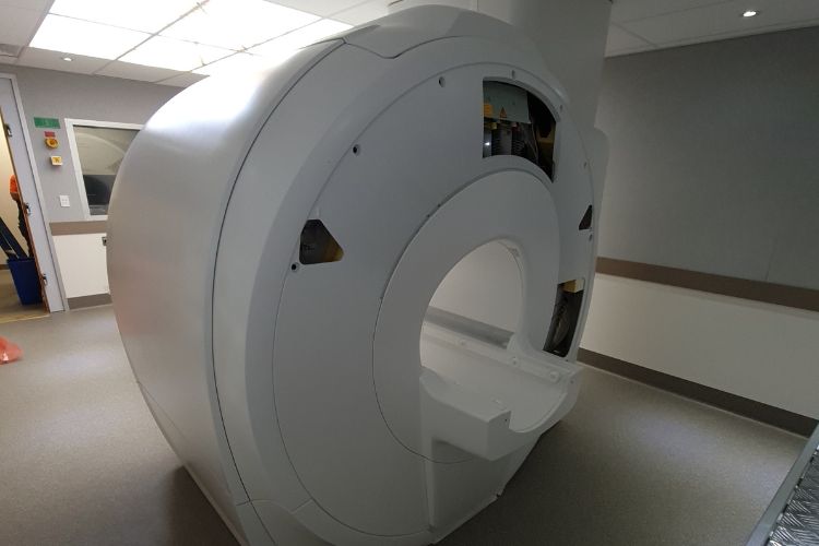
Magnetic Resonance Imaging (or MRI) is a medical imaging technique that uses magnetic fields and radio waves to create detailed images of your body’s organs and tissues, to detect, diagnose and monitor diseases and treatments.

In contrast to X-rays, MRIs are effective at scanning soft tissue. The brain, spinal cord, nerves, muscles, ligaments, and tendons are clearly visualised with this imaging method, and therefore, knee and shoulder injuries are often imaged by an MRI machine. The different matter that make up aneurysms and tumours in the brain can also be observed using MRI technology.
Computed Tomography (CT Scan)
CT scans, or computed tomography, are advanced imaging procedures that use a narrow beam of X-rays rotated around the body. While CT scans expose patients to ionising radiation, the benefits often outweigh the risks due to their diagnostic accuracy and ability to guide effective treatments.
These rays produce signals that are processed digitally to generate cross-sectional images which are then stacked together to create a three-dimensional image of the patient’s body to allow for easy identification of basic structures and tumours or abnormalities.
CT scans are a useful screening tool to identify disease or injury within various regions of the body. For example, medical professionals would suggest having a CT scan to image the head to locate injuries, tumours, and clots leading to stroke or hemorrhage, or to detect possible tumours or lesions within the abdomen.
Likewise, CT scans can capture the lungs to reveal the presence of tumours, pulmonary embolisms (blood clots), excess fluid, emphysema or pneumonia, complex bone fractures, severely eroded joints, and bone tumours that require more detail than an X-ray.
Ultrasound
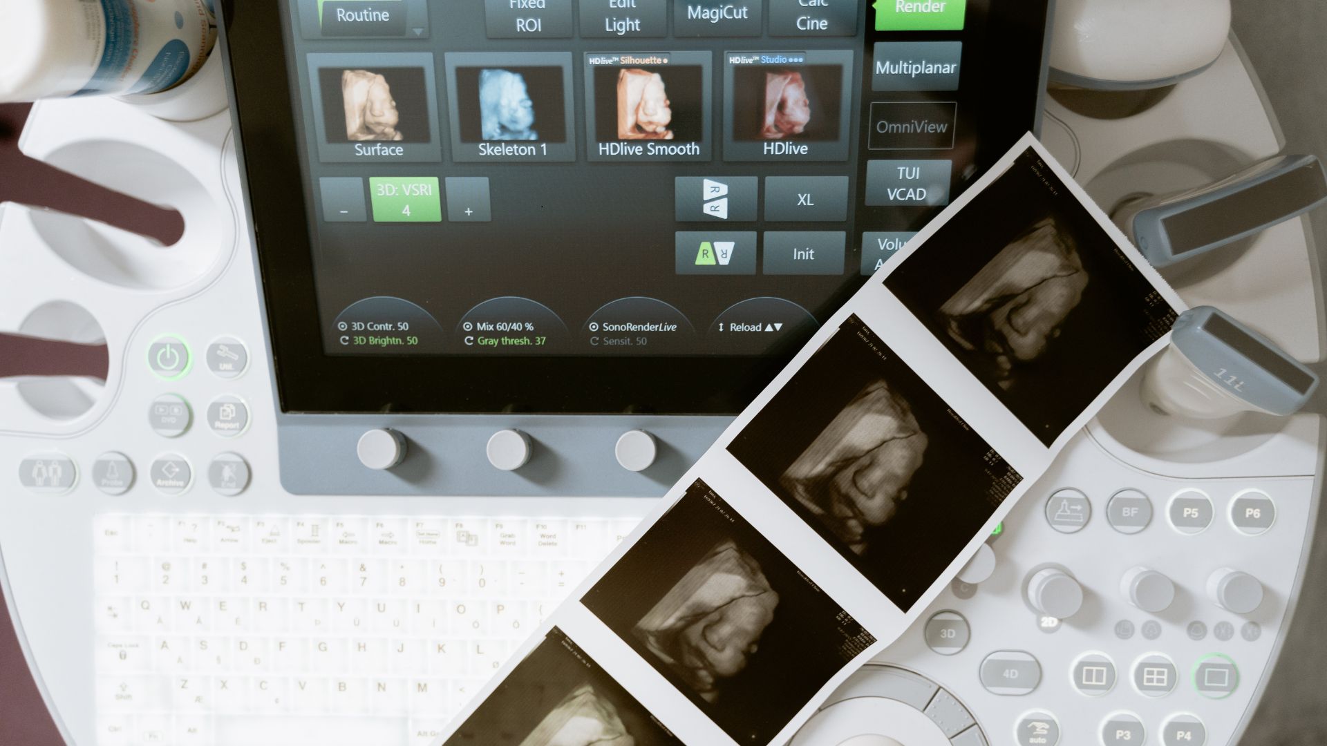
If your doctor requires visualisations of your abdominal and pelvic organs to diagnose abdominal pain, musculoskeletal, cardiac and vascular systems, or to check foetal development during pregnancy, you may be asked to have an ultrasound.
During an ultrasound scan, a device called a transducer is placed on the skin with a special gel that helps transmit sound waves. These sound waves bounce off tissues and organs, creating images displayed on a monitor. In some cases, specialised probes may be inserted into the body to obtain clearer images of internal structures.
When is Medical Imaging Necessary?
Medical imaging is a vital part of healthcare services and has many benefits and purposes, such as:
- Early detection: Medical imaging can assist doctors in detecting health issues before external symptoms appear, or what an external examination may be insufficient to reveal. For example, a mammogram can help detect breast cancer.
- Screening and diagnosis: Imaging tests provide medical professionals with additional information about what symptoms have caused a diagnosis.
- Planning: Imaging tests are used to plan treatment, such as what surgery or radiotherapy will be required.
- Monitoring and assessing: Radiographers will perform imaging tests as the results can be used to monitor the progression of an illness or how well a patient is responding to treatment.
- Improving fetal health: Medical imaging is necessary to monitor a baby’s health and progress in the womb.
Ultimately, consult healthcare providers for imaging needs and what type is suited for your condition.

Frequently Asked Questions About Medical Imaging
What risks are there with medical imaging?
Many patients worry about exposure to radiation from medical imaging procedures. While it’s true that some use small amounts of ionising radiation, the health risks are low. These imaging technologies have been tested for safety, are conducted by trained radiology professionals and are subject to strict government and industry regulations. Ultimately, it’s important to remember they are extremely beneficial in achieving the right diagnosis.
What are the costs involved with medical imaging?
Medical imaging can be expensive. At PRP Diagnostic Imaging, Medicare covers a wide range of our services, though, for certain examinations and procedures, there may be an out-of-pocket fee or a ‘gap fee.’ Nonetheless, we ensure all patients know the full cost of the examination you require before its commencement.
How do I prepare for my diagnostic imaging procedure?
Our clinics at PRP Diagnostic Imaging are doctor-owned and operated. Our priority is the high-quality care of our patients.
Each procedure is different, so you need to pay attention to the specific instructions you’ll receive beforehand. When you make an appointment, our helpful staff will explain clearly what you need to do to prepare. Please don’t hesitate to contact us with any questions you have.
Have a referral from your doctor for a scan? Send us a request form with your referral, and we’ll be in touch about booking you an appointment in no time. Located all across NSW, PRP Diagnostic Imaging offers all types of medical imaging from ultrasounds, x-rays, MRIs, CT scans and more.
